Excitable Tissue: Muscle

Introduction
Muscle cells, like neurons, can be excited chemically, electrically, and mechanically to produce an action potential that is transmitted along their cell membranes. Unlike neurons, they respond to stimuli by activating a contractile mechanism. The contractile protein myosin and the cytoskeletal protein actin are abundant in muscle, where they are the primary structural components that bring about contraction.
Skeletal muscle morphology organization
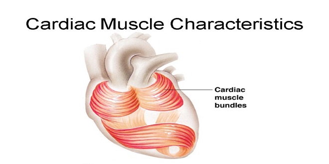
Skeletal muscle is made up of individual muscle fibers that are the “building blocks” of the muscular system in the same sense that the neurons are the building blocks of the nervous system. Most skeletal muscles begin and end in tendons, and the muscle fibers are arranged in parallel between the tendinous ends, and muscle fibers, and is generally under voluntary control. Cardiac muscle also has cross-striations, but it is functionally syncytial and, although it can be modulated via the autonomic nervous system, it can contract rhythmically in the absence of external innervation owing to the presence in the myocardium of pacemaker cells that discharge spontaneously.
Dystrophin–glycoprotein complex
The large dystrophin protein (molecular mass 427,000 Da) forms a rod that connects the thin actin filaments to the transmembrane protein β-dystroglycan in the sarcolemma by smaller proteins in the cytoplasm, syntrophins. β-dystroglycan is connected to merosin (me rosin refers to laminas that contain the α2 subunit in their trimetric makeup) in the extracellular matrix by α-dystroglycan.
Sarcotubular system
The muscle fibrils are surrounded by structures made up of membranes that appear in electron photomicrographs as vesicles and tubules. These structures form the sarcotubular system, which is made up of a T system and sarcoplasmic reticulum. The T system of transverse tubules, which is continuous with the sarcolemma of the muscle fiber, forms a grid perforated by the individual muscle fibrils.
Ion distribution & fluxes
The distribution of ions across the muscle fiber membrane is similar to that across the nerve cell membrane. Approximate values for the various ions and their equilibrium potential. As in nerves, depolarization is largely a manifestation of Na+ influx, and repolarization is largely a manifestation of K+ efflux.
The muscle twitch
A single action potential causes a brief contraction followed by relaxation. This response is called a muscle twitch, the action potential and the twitch are plotted on the same time scale. The twitch starts about 2 ms after the start of depolarization of the membrane before repolarization is complete. The duration of the twitch varies with the type of muscle being tested. “Fast” muscle fibers, primarily those concerned with fine, rapid, precise movement, have twitch durations as short as 7.5 ms. “Slow” muscle fibers, principally those involved in strong, gross, sustained movements, have twitch durations up to 100 ms.
Summation of contractions
The electrical response of a muscle fiber to repeated stimulation is like that of a nerve. The fiber is electrically refractory only during the rising phase and part of the falling phase of the spike potential. At this time, the contraction initiated by the first stimulus is just beginning. However, because the contractile mechanism does not have a refractory period, repeated stimulation before relaxation has occurred produces additional activation of the contractile elements and a response that is added to the contraction already present.
Summary
Skeletal muscle is a true syncytium under voluntary control. Skeletal muscles receive electrical stimuli from neurons to elicit contraction: “excitation-contraction coupling.” Action potentials in muscle cells are developed largely through the coordination of Na+, K+, and Ca2+ channels. Contraction in skeletal muscle cells is coordinated through Ca2+ regulation of the act myosin system that gives the muscle its classic striated pattern under the microscope.


