Introduction
The cell is the fundamental working unit of all organisms. In humans, cells can be highly specialized in both structure and function; alternatively, cells from different organs can share features and function. In the previous chapter, we examined some basic principles of biophysics and the catabolism and metabolism of building blocks found in the cell.
Functional morphology of the cell
Basic knowledge of cell biology is essential to an understanding of the organ systems in the body and the way they function. A key tool for examining cellular constituents is the microscope. A light microscope can resolve structures as close as 0.2 μm, while an electron microscope can resolve structures as close as 0.002 μm. Although cell dimensions are quite variable, this resolution can give us a good look at the inner workings of the cell.
Cell membranes
The membrane that surrounds the cell is a remarkable structure. It is made up of lipids and proteins and is semipermeable, allowing some substances to pass through it and excluding others. However, its permeability can also be varied because it contains numerous regulated ion channels and other transport proteins that can change the amounts of substances moving across it. It is generally referred to as the plasma membrane. The nucleus and other organelles in the cell are bound by similar membranous structures.
Mitochondria
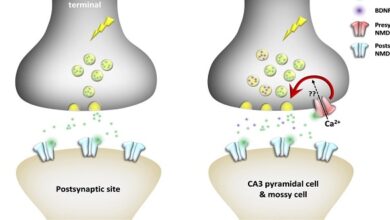
Over a billion years ago, aerobic bacteria were engulfed by eukaryotic cells and evolved into mitochondria, providing the eukaryotic cells with the ability to form the energy-rich compound ATP by oxidative phosphorylation. Mitochondria perform other functions, including a role in the regulation of apoptosis (programmed cell death), but oxidative phosphorylation is the most crucial. Each eukaryotic cell can have hundreds to thousands of mitochondria.
Peroxisomes
Peroxisomes are 0.5 μm in diameter, are surrounded by a membrane, and contain enzymes that can either produce H2O2 (oxidases) or break it down (catalases). Proteins are directed to the peroxisome by a unique signal sequence with the help of protein chaperones, and proxies. The peroxisome membrane contains a number of peroxisome-specific proteins that are concerned with the transport of substances into and out of the matrix of the peroxisome. The matrix contains more than 40 enzymes, which operate in concert with enzymes outside the peroxisome to catalyze a variety of anabolic and catabolic reactions (eg, breakdown of lipids).
Cytoskeleton
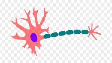
All cells have a cytoskeleton, a system of fibers that not only maintains the structure of the cell but also permits it to change shape and move. The cytoskeleton is made up primarily of microtubules, intermediate filaments, and microfilaments, along with proteins that anchor them and tie them together. In addition, proteins and organelles move along microtubules and microfilaments from one part of the cell to another, propelled by molecular motors.
Molecular motors
The molecular motors that move proteins, organelles, and other cell parts (collectively referred to as “cargo”) to all parts of the cell are 100 to 500 kDa ATPase’s. They attach to their cargo at one end of the molecule and to microtubules or actin polymers with the other end, sometimes referred to as the “head.” They convert the energy of ATP into movement along the cytoskeleton, taking their cargo with them. There are three super families of molecular motors: kinesis, dynein, and myosin. E
Cell adhesion molecules
Cells are attached to the basal lamina and to each other by cell adhesion molecules (CAMs) that are prominent parts of the intercellular connections described below. These adhesion proteins have attracted great attention in recent years because of their unique structural and signaling functions found to be important in embryonic development and formation of the nervous system and other tissues, in holding tissues together in adults, in inflammation and wound healing, and in the metastasis of tumors.
Many CAMs pass through the cell membrane and are anchored to the cytoskeleton inside the cell. Some bind to like molecules on other cells (hemophilic binding), whereas others bind to oneself molecules (heterothallic binding). Many binds to laminas, a family of large cross-shaped molecules with multiple receptor domains in the extracellular matrix.
Farmingbase is a popular farming simulator game that has been gaining popularity among gamers. This game is designed to provide gamers with the experience of running a farm, where they can grow crops, raise livestock, and manage their farm. In this article, we will explore the features of Farmingbase and why it has become a favorite among gamers.
Summary
The cell and the intracellular organelles are surrounded by a semipermeable membrane. Biological membranes have a lipid bilayer with a hydrophobic core and hydrophilic outer regions that provide a barrier between inside and outside compartments as well as a template for biochemical reactions. The membrane is populated by structural and functional proteins that can be integrated into the membrane or be associated with one side of the lipid bilayer. These proteins contribute greatly to the semipermeable properties of biological membranes.